

Posted by: Central Florida Eye Specialists in Uncategorized

The human eye is one crucial aspect that we should not take for granted. The miracle of seeing the beautiful world around you is the greatest gift we have as individuals. The uniqueness of these mysterious organs is the reason that eyes make all magical experiences through sight. Did you know that the human eye can differentiate approximately 10 million different colors? The eye anatomy is very intricate and essential to understanding how our eyes can manage the value they bring to our lives. Here is a journey of the eye anatomy, starting from the front and working to the back of the eye.
Outside of the Eye
The eye sits in a protective bony socket called the orbit. Six extraocular muscles in the orbit that is attached to the eye. These muscles move the eye up and down, side to side, and rotate the eye.
The extraocular muscles are attached to the white part of the eye called the sclera. This part of the eye is a strong layer of tissue covering nearly the entire surface of the eyeball.

The Surface of the Eye
The eye’s surface and the inner surface of the eyelids are covered with a transparent membrane called the conjunctiva.
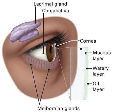
Tears lubricate the eye and makeup three layers. These three layers together are called the tear film. The conjunctiva makes the mucous layer. The lacrimal gland, the watery part of the tears, is produced. The eye’s lacrimal gland sits under the outside edge of the eyebrow (away from the nose) in the orbit. The meibomian gland makes the oil that becomes another part of the tear film. Tears drain from the eye through the tear duct.
Front of The Eye
Light is focused into the eye through the clear, dome-shaped front portion of the eye called the cornea. Behind the cornea is a fluid-filled space called the anterior chamber. The fluid is called aqueous humor. The eye is consistently producing aqueous humor. Aqueous humor also drains from the eye in an area called the drainage angle to maintain constant eye pressure.